Surface Plasmon Resonance Biosensors
The impact
Diabetes affects around 5% of the world’s population and is associated with a wide range of health problems, including heart disease, strokes, kidney disease, eye problems, and death. While diabetes cannot be cured, its effects can be controlled if properly managed. The glucose concentration in human blood is most commonly measured using a finger-pricking invasive testing device. However, these devices may cause patient discomfort and introduce a risk of infection. Accordingly, the development of noninvasive (NI) testing devices has attracted increasing interest in recent years.
In OPSENLAB, a novel technique for NI glucose monitoring is developed based on Polarimetry-Surface Plasmon Resonance (SPR) prism coupler with a resolution of 10 mg/dL. In general, the proposed technique provides reliable and significant potential approach for noninvasive glucose monitoring device (NIGMD) applications.
The measurement system
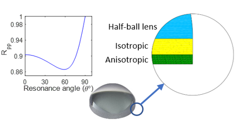
SPR prism coupler: consists of a half-ball glass lens, a Cr-Au isotropic thin-film layer and a Ta2O5 anisotropic. Total internal reflection (TIR) is generated at the sensed surface and SPR is induced. The change in glucose concentration results in changing refractive index of sample thus will be sensed by SPR sensor.
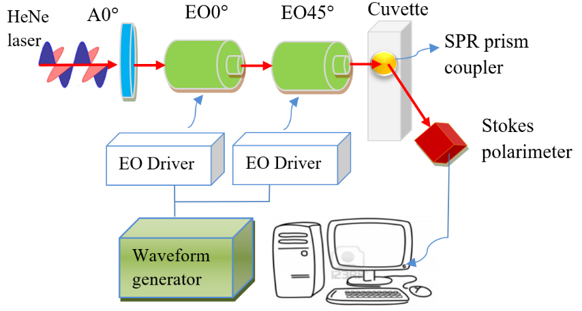
Dynamic polarization scanning ellipsometry measurement system is built for extracting the glucose concentration enhanced by SPR prism coupler.
The results
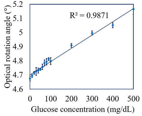
The optical rotation angle increases linearly with the glucose concentration of tissue phantom solution over the range of 0~500 mg/dL with a correlation coefficient of R2=0.9871.
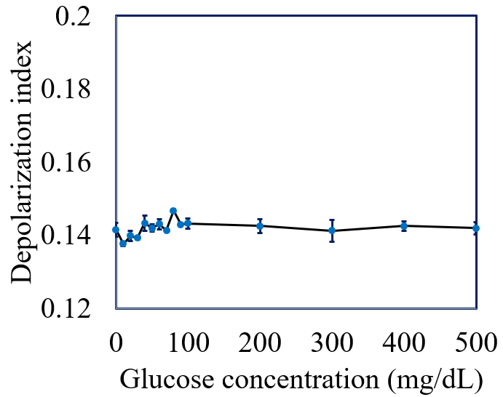
The depolarization index has a constant value of approximately 0.14 irrespective of the glucose concentration due to the TIR effect produced by the SPR coupler, which minimizes the depolarization.
The pilot study: Oral Glucose Tolerance Tests (OGTTs)
Oral glucose tolerance tests (OGTTs) were performed on four healthy human volunteers aged between 20 and 25 years old. All of the subjects were male and Asian. The subjects were asked to fast for 8 hours and the blood glucose concentration was then measured by pressing the fingertip against the flat surface of the SPR prism coupler. The volunteers then drank 750 ml of aqueous solution containing 75 g sugar and the glucose measurement process was performed once again after an interval of 2 hours. The extracted values of the optical rotation angle increased following glucose ingestion for all of the volunteers. In other words, the results are consistent with the measurement results obtained for tissue phantom solutions.
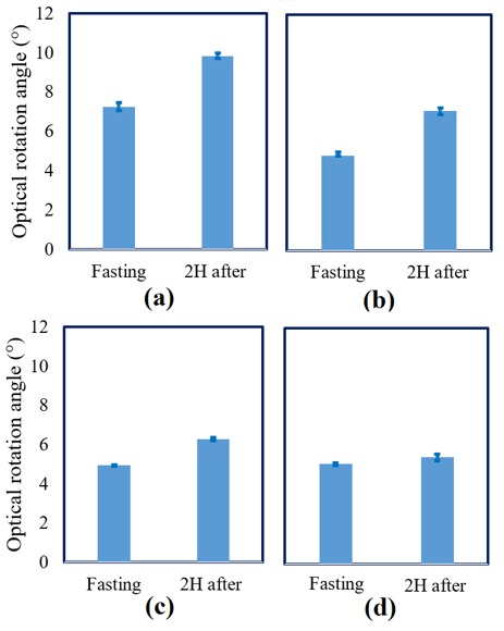
Extracted values of optical rotation angle in OGTTs pilot study for:
(a) subject #1, (b) subject #2, (c) subject #3, and (d) subject #4.