Noninvasive Glucose Sensing
The impact
Diabetes is a major healthcare concern nowadays; with a rapidly growing incidence worldwide and massive economic implications. Accordingly, many techniques have been proposed for detecting diabetes by measuring the glucose level in human blood, classified as either invasive, minimal invasive, or non-invasive (NI), depending on the transduction mechanism used. Invasive methods are the most commonly used due to their simplicity and accuracy. However, invasive methods are often rather painful and carry an increased risk of infection. While minimal invasive techniques are expensive and require complicated calibration procedures. As a result, NI techniques became a potential tool for glucose concentration detection in the future.
In OPSENLAB, a novel technique for NI glucose monitoring is developed based on Stokes-Mueller matrix polarimetry with a resolution of 10 mg/dL. In general, the proposed technique provides reliable and significant potential approach for noninvasive glucose monitoring device.
The methodology
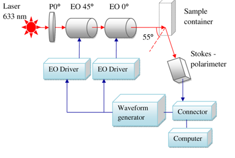
Differential Mueller matrix formalism is developed for circular birefringence property detection of turbid media. Dynamic polarization scanning ellipsometry measurement system is built for extracting optical rotation angle and depolarization index parameters, and consequently the glucose concentration. The dynamic polarization scanning ellipsometry consisting of a polarization scanning generator (PSG) and a high-accuracy Stokes polarimeter is used to sense the glucose concentration in aqueous solutions, respectively.
The results
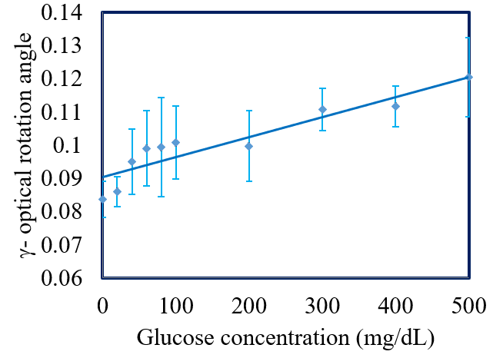
The optical rotation angle increases linearly with an increasing glucose concentration. The standard deviation of the experimental values obtained over four repeated tests for each glucose sample was found to be 8.410-3 degree and the sensitivity were determined to be 610-5deg./(mg/dL).
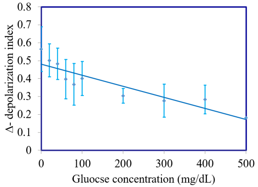
Depolarization index () decreases linearly with an increasing glucose concentration with a standard deviation of 8.010-2 and the sensitivity of the depolarization index measurements was determined to be 610-5/ (mg/dL).
The pilot study
A further series of experiments was performed to measure the optical rotation angle and depolarization index of the extracellular tissue on the human fingertip of three healthy volunteers at three different periods of the day defined relative to the standard meal times, i.e., breakfast (around 9.00), lunch (around 12.00) and dinner (around 17.00).
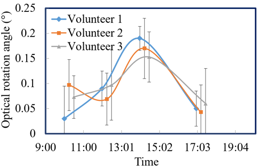
The optical rotation angle values increase after meal times (i.e., 10:00 and 14:00), but decrease before meal times (i.e., 12:00 and 17:00).
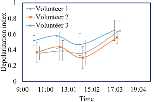
The depolarization index increases before meal times (i.e., 12:00 and 17:00), but decreases after meal times (i.e., 10:00 and 14:00)